
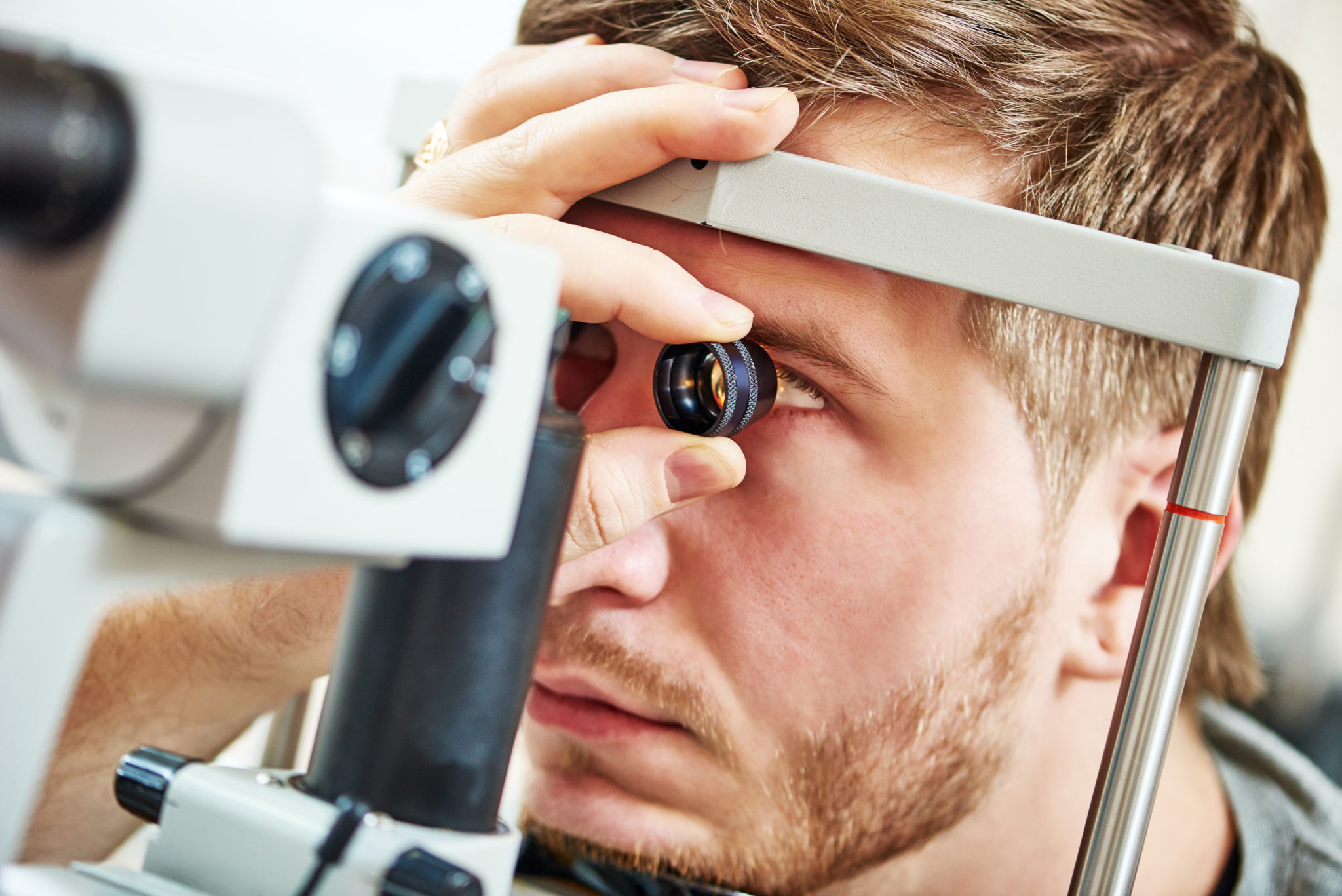
Ultra-widefield photography
This diagnostic tool captures a 200-degree panoramic view of the back of the eye in a single scan, showing the vast majority of the retina. This technology allows for the simultaneous evaluation of central and peripheral eye health, which traditional, narrower viewing methods miss. Images can be magnified to examine fine details, while specialized color filters isolate different retinal layers to spot hidden pathologies. This detailed analysis is vital for detecting macular degeneration, diabetic retinopathy complications, vascular changes from high blood pressure, optic nerve variations, ocular tumors, and peripheral retinal problems like holes, tears, or detachments.
How Optomap helps
The same scan supports some of the most important medical exams we do.
Diabetic eye examsThis technology allows for the simultaneous evaluation of central and peripheral blood vessels, which traditional, narrower viewing methods miss. Specialized color filters isolate different retinal layers to spot hidden diabetic retinopathy complications and peripheral vascular leaks. Diabetic eye exams
Macular degenerationHigh-resolution digital imaging used to track macular changes and monitor both dry and wet age-related macular degeneration. The digital maps allow for direct, long-term comparisons of the retina to evaluate the progression of geographic atrophy and document central drusen distribution. Macular degeneration care
Related care
Diagnostic testing
We utilize state-of-the-art technology to evaluate and diagnose sight-threatening conditions like macular degeneration, glaucoma, diabetic retinopathy, and other retinal complications.
Glaucoma
Glaucoma is a group of eye diseases that cause progressive, irreversible damage to the optic nerve head and the underlying retinal cell layers.
Cataracts
A cataract is the gradual clouding of the eye’s natural crystalline lens, which sits behind the pupil and functions like a camera lens to focus light onto the retina.

Ask about an Optomap scan
Call our Safford office to add Optomap imaging to your next eye exam.